In the novel Water Margin, one of the Four Great Classical Novels in China, there is one chapter Wu Song Fights Tiger, in which Wu Song’s reaction to the suddenly-appeared tiger is vividly described. The verbs, such as “immediately turned”, “dodged”, “evaded”, “jumped”, “grabbed”, etc. have perfectly shown Wu Song’s psychological changes and physiological responses when facing threat and danger. In psychology, it is defined as fight-or-flight response, namely, the body’s response to perceived threat or danger, which was originally named for its ability to enable people to physically fight or run away when faced with danger. During this process, certain hormones, such as adrenalin and cortisol, are released, speeding the heart rate, slowing digestion, shunting blood flow to major muscle groups, and changing various other autonomic nervous functions, giving the body a burst of energy and strength. When the perceived threat is gone, systems are designed to return to normal function via the relaxation response. As we all know, when in danger, it’s natural to feel afraid. This fear triggers many split-second changes in the body to prepare to defend against the danger or to avoid it. This “fight-or-flight” response is a healthy reaction meant to protect a person from harm.
However, if people suffered excessively or repeatedly from fight-or-flight responses, it is bound to damage their health, which is defined as post-traumatic stress disorder (PTSD). People who suffered from PTSD may feel stressed or frightened even when they’re no longer in danger. PTSD was first brought to public attention in relation to war veterans, but it can result from a variety of traumatic incidents, such as mugging, rape, torture, being kidnapped or held captive, child abuse, car accidents, train wrecks, plane crashes, bombings, or natural disasters such as floods or earthquakes. Nowadays, PTSD has grown to be one of the hot issues in brain science. It is believed that the exploration of the neural circuits causing fight-or-flight response may be the source of PTSD pathogenesis. Yet little is known about the neural circuits specifically processing threat-relevant sensory information in the mammalian brain.
In a new study published on 26th June 2015 by Science, a research group led by Professor Peng Cao of Institute of Biophysics, Chinese Academy of Sciences, made great progress in parvalbumin-positive excitatory visual pathway to trigger fight-or-flight response in mice.
By making use of an optogenetic approach, the research team found that activation of neurons expressing channelrhodopsin-2 (ChR2) in mouse superior colliculus caused freezing, which encouraged their systematical identification of the key neuronal subtypes underlying that behavior.
In their study, it is identified that parvalbumin-positive (PV+) excitatory projection neurons in mouse superior colliculusis(SC) a key neuronal subtype for detecting looming objects and triggering fight-or-flight response. These neurons, which are distributed mainly in the superficial superior colliculus (SC), divergently project to different brain areas, including the parabigeminal nucleus (PBG), namely, an intermediate station leading to the amygdala. Activation of the PV+ SC-PBG pathway caused fight-or-flight response, induced conditioned aversion, and resulted in depression-like behaviors.
The authors conclude that the SC PV+ neurons forms a subcortical visual pathway that transmits threat-related visual information to the amygdala to cause fear responses, together with earlier studies, suggesting a “retina-SC-PBGN-amygdala hypothalamus” pathway for vision-induced fear responses. In addition, the SC PV+ neurons in the superficial gray layer are predominantly glutamatergic projection neurons with spiking patterns distinct from those of their counter parts in cortical regions, broadening the concept of PV+ neurons and adding another perspective to understanding their functions. Last but not least, the SC PV+ neurons may belong to type-ρ looming detector, supporting the notion that mathematically defined computational units correspond to specific neuronal subtypes. Those outcomes lay a solid theoretical foundation for further study of PTSD pathogenesis.
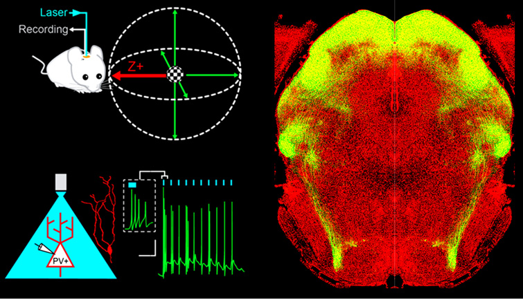
Fig. A, PV+ neurons detect soccer ball-like looming stimulus in the visual field. B, The morphological and physiological properties of PV+ neurons in SC. C, The downstream pathways formed by PV+ neurons in SC (from Science Magzine)