
The Center of Bio-Imaging develops a new type of supporting film covering on TEM grid used for single-particle cryo-electron microscopy-amorphous nickel titanium alloy film
The Center of Bio-Imaging of the Institute of Biophysics, the SUN Fei's group of the National Laboratory of Biomacromolecules, and the YIN Changcheng's group of Peking University jointly published an article entitled "Amorphous nickel titanium alloy film: A new choice for cryo electron microscopy sample preparation" on Progress in Biophysics and Molecular Biology on August 6, 2020. In the research, nickel-titanium alloy materials were used to prepare supporting film covering on TEM grid used for single-particle cryo-electron microscopy.
Single-particle cryo-electron microscopy is one of the important tools for structural biology research. The sample type targeted by this technique is a biomacromolecule solution. Compared with X-ray crystallography, this technique has low requirements on samples and does not require crystallization, but there are still many bottlenecks in sample preparation. An ideal cryo-electron microscopy sample requires the sample to be uniformly dispersed in a complete form and randomly distributed in the thin ice layer in the grid hole, and the sample to have sufficient conductivity. However, in the real-life conditions, sample particles are mainly distributed on the grid hole instead of in the grid hole, the sample amount distributed in the hole is small, the sample distribution has an orientation advantage, and the sample is denatured under the action of the air-water interface, poor conductivity of the sample leads to serious image drift of cryo-electron microscopy. Existing solutions for these problems include laying continuous ultra-thin films (carbon film, graphene film, graphene oxide film, protein two-dimensional crystal film, etc.), using affinity membranes to selectively absorb more samples onto the continuous ultra-thin film, and changing the material of the supporting film (such as using pure gold supporting film) and so on. These solutions can improve one or more problems, or make the sample evenly distributed in the grid hole, or keep the sample away from the air-water interface to avoid orientation advantages and sample denaturation, or improve the conductivity of the overall sample by enhancing the conductivity of the supporting film, etc.
In this achievement, the researchers used nickel-titanium alloy to prepare amorphous nickel titanium alloy film (Figure 1). Unlike the supporting film made of pure gold, this nickel titanium supporting film and the traditional carbon microarray supporting film are both amorphous and have the same the usage method, so it can be directly applied to the cryo-electron microscopy sample preparation and data collection. The researchers found that compared with the traditional carbon microarray supporting film, the nickel titanium alloy one has better conductivity (4 orders of magnitude higher), which can effectively reduce the sample drift caused by electron beam irradiation. Under the same conditions, a higher resolution cryo-electron microscopy three-dimensional reconstruction result can be obtained. In addition, the surface properties of the amorphous nickel titanium alloy film are different from that of the traditional carbon film, which means biomolecules have very small non-specific interaction with the amorphous nickel titanium alloy film (one order of magnitude smaller than that with the traditional carbon film). This prevents the biological sample from being deformed and focused due to the non-specific interaction with the supporting film, so the distribution density in the amorphous nickel titanium alloy film hole is higher and the dispersion is better. Besides, cell culture experiments show that the amorphous nickel titanium alloy film has low biological toxicity and can be used for cell growth and in-situ cryo-electron microscopy studies. These experimental results show that the amorphous nickel titanium alloy film is an excellent grid for single-particle cryo-electron microscopy, providing a new choice for cryo-electron microscopy researchers.
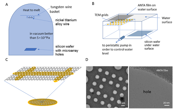
Figure 1. Preparation method and cryo-electron microscope photograph of amorphous nickel titanium alloy film covering on TEM grid
(Image by Dr. SUN Fei's group)
The research results were published on Progress in Biophysics and Molecular Biology at the invitation of Professor Wah Chiu, editor-in-chief of Stanford University and Professor Sheemei Lok of the National University of Singapore, as part of the special issue CryoEM Developments.
A Chinese invention patent was applied for the research results (application number 201810326897.5) and technology authorization was given in 2018. The results were authorized to Zhenjiang Lehua Electronic Technology Co., Ltd. for production and sales (trade name "CryoMatrix-Amorphous Alloy Film"), providing services for cryo-electron microscopy researches in China. At present, many users have used the supporting film to prepare cryo-electron microscopy samples. Representative results include:
(1) Hua, T. et al. (2020). Activation and Signaling Mechanism Revealed by Cannabinoid Receptor-Gi Complex Structures, Cell. 180, 655-665 e18.
(2) Qiao, A. et al. (2020). Structural basis of Gs and Gi recognition by the human glucagon receptor. Science, 367: 1346-1352.
(3) Wu, C. et al. (2019). High-quality, high-throughput cryo-electron microscopy data collection via beam tilt and astigmatism-free beam-image shift. J. Struct. Biol. 208, 107396.
The correspondence author of this article is SUN Fei, a researcher at the National Laboratory of Biomacromolecules. HUANG Xiaojun, a professor of engineering from the Protein Platform Bio-Imaging Center, and Dr. ZHANG Lei from YIN's group of Peking University School of Medicine, are the co-first authors of this article. WEN Zuoling from SUN's group participated in cryo-electron microscopy image processing, CHEN Hui of LOU Jizhong's group participated in atomic force measurement experiments, and LI Shuoguo and JI Gang from the Center of Bio-Imaging participated in cell culture and fluorescence experiments. Thanks to YAN Xiyun's group for providing high-quality ferritin as a test sample, and to Dr. DING Wei from the Institute of Physics for his help in the preliminary data processing. The research has been funded by the National Natural Science Foundation of China, the key research and development projects of the Ministry of Science and Technology, the Key Technological Innovation Talent Fund of the Chinese Academy of Sciences and the Beijing Municipal Science and Technology Commission.
The web link of this paper: https://www.sciencedirect.com/science/article/pii/S0079610720300742
Contact: SUN Fei
Institute of Biophysics, Chinese Academy of Sciences
Beijing 100101, China
Email: feisun@ibp.ac.cn
(Reported by Dr. SUN Fei's group)