
Center for Biological Imaging of Core Facilities for Protein Science and the collaborators of Sino-Danish College have made new breakthroughs in the field of volume microscopy for biological samples
September 2, 2021, an article entitled " Cellular 3D-reconstruction and analysis in human cerebral cortex using automatic serial sections" was published on-line in Communications Biology. Based on the automatic collector of ultrathin sections scanning electron microscopy (AutoCUTS-SEM, Journal of Structural Biology, 200 (2017) 87-96)" technology, the Automatic Collector of serial sections for Light microscopy (AutoCUTS-LM) was developed in this study for pyramidal neurons in three-dimensional (3D) reconstruction from old archived human brain tissue. Through 3D reconstruction and analysis of tissue structure, this technique provided a better understanding of changes in molecules and function. The results revealed the detailed structure of pyramidal cells (number, volume, diameter, sphericity and orientation) and their 3D spatial organization were arranged in a columnar structure.
The workflow in this study involved (1) advanced tissue sampling methods of cerebral cortex, (2) an automated serial section collection system, (3) digital tissue library, (4) cell detection using convolution neural network, (5) 3D cell reconstruction, and (6) advanced analysis. For light microscopic imaging, special attention was paid to the flatness of sections, transmittance of collecting tape and dyeing intensity. Serial sections (>200nm thickness) were extremely prone to wrinkles, which affected the image quality. This study systematically compared the effects of different section thickness, cut speed, ambient temperature and humidity, material and hydrophilic treatment conditions of the collection tape, and the fixed method of tape on the flatness of the sections. The effects of dyeing solution concentration, dyeing time, temperature and method on image contrast were also compared. Through these efforts, the wrinkles of the sections have been eliminated.
Compared with the AutoCUTS-SEM, AutoCUTS-LM developed in this study greatly shortens the period of sample preparation and data collection. The research cost has also been significantly reduced, which makes larger-scale biological samples easier to study. In addition, the pipeline of these combined techniques provides a detailed analysis of tissues and cells in biology and pathology.
In the collaborative study, Prof. SUN Fei at Institute of Biophysics, CAS, and Prof. Jens R. Nyengaard at Aarhus University co-direct this work. Nick Y. Larsen at Aarhus University and Sino-Danish College, University of CAS is the first authors. Dr. LI Xixia, Dr. JI Gang and engineer assistant TAN Xueke at Center for Biological Imaging of Core Facilities for Protein Science have made substantial contributions to this research in sample preparation, automatic collection of continuous sections and image acquisition.
The work was supported by grants from National Natural Science Foundation of China (31925026) and the Chinese Academy of Sciences (QYZDB-SSW-SMC004).
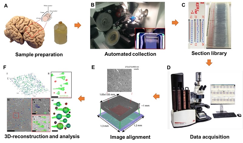
Fig1.The pipeline of AutoCUTS-LM
Article link: https://www.nature.com/articles/s42003-021-02548-6
Contact: Sun Fei
Institute of Biophysics, Chinese Academy of Sciences
Beijing 100101, China
Email: feisun@ibp.ac.cn
(Reported by Center for Biological Imaging)