
Cortical Basis of the Human Saliency Map
Why do our eyes instinctively jump to a bright red apple on a green tree or a flashing light in the dark? The brain's ability to detect and prioritize visually "salient" stimuli-those that stand out from their surroundings-is essential for survival and efficient perception. Yet, for decades, neuroscientists have debated where in the brain these saliency signals originate.
In a study published in PLoS Biology on October 14, 2025, a research team led by Professor ZHANG Peng from the Institute of Biophysics of the Chinese Academy of Sciences, employed vascular space occupancy (VASO)-based laminar fMRI with ultra-high spatial specificity at 7 Tesla (VASO laminar fMRI) to uncover the cortical microcircuit mechanisms underlying the human bottom-up attentional saliency map.
The researchers discovered that the bottom-up attention saliency map originates from lateral inhibition within the superficial layers of the primary visual cortex (V1) and is transmitted through feedforward pathways to the intraparietal sulcus (IPS), thereby enabling efficient information selection.
In the experiments, the visual stimuli consisted of background bars of uniform orientation and a small patch of foreground bars with a different orientation. The visual saliency of the foreground was precisely manipulated by varying the orientation difference between the foreground and background.
Behavioral results showed that participants exhibited better detection performance for foreground stimuli with higher visual saliency.
The submillimeter-resolution 7T VASO fMRI data revealed that the superficial layers of V1 encoded the visual saliency map, consistent with a laminar pattern of lateral connections, and that this signal was fed forward to the middle layers of the IPS.
Moreover, the strength of the saliency signal in the superficial layers of V1 accurately predicted participants' behavioral detection performance for the salient target.
These findings support the notion that the visual saliency map is computed within the superficial layers of V1 via lateral connections and transmitted to higher cortical regions, such as the parietal cortex, to facilitate bottom-up attentional selection.
This study provides crucial mesoscale functional evidence in the human brain supporting the "V1 Saliency Hypothesis" of visual attention computation and may contribute to the development of brain-inspired visual models.
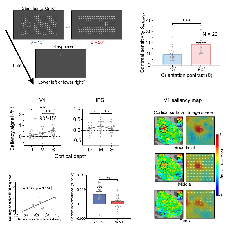
Figure: Generation and transmission mechanisms of the bottom-up attentional saliency map
(Image by ZHANG Peng's group)
Article link: https://doi.org/10.1371/journal.pbio.3003159
Contact: ZHANG Peng
Institute of Biophysics, Chinese Academy of Sciences
Beijing 100101, China
E-mail: zhangpeng@ibp.ac.cn
(Image by Prof. ZHANG Peng's group)