Sarcomeres Pattern Proprioceptive Sensory Dendritic Endings through UNC-52/Perlecan in C. elegans
Sensory dendrite morphogenesis is a critical step to establish the receptor fields of neurons and to assembly functional neural circuits. In the vertebrate proprioceptive system, sensory terminals wrap around specialized muscle fibers to form the encapsulated sensory receptors: the muscle spindles. For proprioceptive neurons, little is known about how the molecular mechanisms that mediate the muscle-nerve interaction.
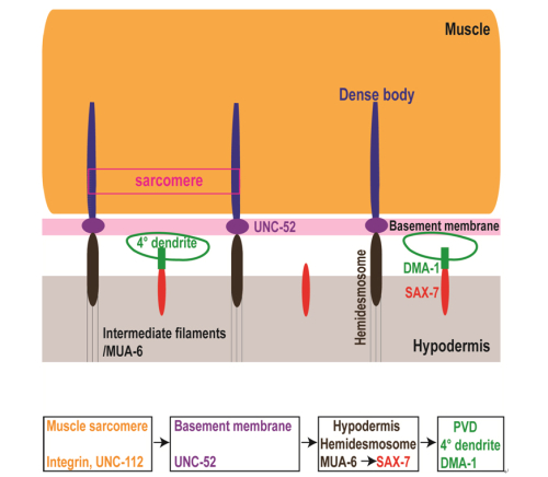
Recently, a PH. D. student Xing liang, Dr. Xiangming Wang and their colleagues under the supervision of professor Kang Shen from the Institute of Biophysics, Chinese Academy of Sciences, investigated the sensory neuron PVD quaternary dendrite morphogenesis in C. elegans. The study finds that the muscularcontractile apparatus provides instructive cues for patterningthe branching of sensory dendrites. The regular pattern ofsarcomere dense bodies, via the ECMprotein UNC-52/Perlecan, defines a SAX-7/L1CAM striped pattern, which in turninstructs the dendritic branch pattern.
The best-understood mammalian proprioceptive receptor is the muscle spindle. Similar to the hypodermal-muscle-dendrite complex in PVD, the muscle spindle contains the sheath cells, specialized muscle fibers and nerve terminals. The sensory nerve terminals wrap around the muscle cells several rounds with evenly spaced turns. Remarkably, in both worms and mammals, the sensory nerves are oriented perpendicular to longitudinal axis of the sarcomeres, a configuration that might maximize the stretching effects from the muscle onto the nerve. Future studies are needed to test if the cellular and molecular interactions that we described here play a similar role in patterning the muscle spindles.
Fig. Schematics of a cross section of the muscle basement membrane, PVD and hypodermis. The MUA-6 is part of the hemidesmosome. Green circles represent cross sections of PVD 4° branches. The signal flow patterning PVD 4° dendrites is muscle/basement membrane/hypodermis/PVD. Each rectangle represents a cell type or tissue. (Liang et al. 2015, Dev. Cell)
This work entitled “Sarcomeres precisely pattern the proprioceptive sensory dendritic endings through Perlecan/UNC-52 in C. elegans” was published online in Developmental Cell on May 14th. It was funded by the National Key Basic Research Foundation of China, the National Natural Science Foundation of China, the Chinese Academy of Sciences, and HHMI.


