
ZHANG Peng Team Publishes Paper on Attention Modulation Mechanism in Human Early Visual Cortex
Progress in Neurobiology published online on August 17, 2020 the latest research by ZHANG Peng's group of the State Key Laboratory of Brain and Cognitive Science of Institute of Biophysics (IBP) of the Chinese Academy of Sciences, titled "Layer-dependent multiplicative effects of spatial attention on contrast responses in human early visual cortex". For the purpose of this research, with support of the IBP's Brain Imaging MRI Platform, the project team was the first in China to set up high-resolution fMRI at 7T and apply the technology to human brain cognitive research.
Using submillimeter-resolution fMRI for multi-modal layering, researchers investigated the effects of top-down spatial attention on the contrast responses across different cortical depths in human early visual cortex. Gradient echo (GE) T2* weighted BOLD signal showed an additive effect of attention on contrast responses across cortical depths. Compared to the middle cortical depth, attention modulation was stronger in the superficial and deep depths of V1, and also stronger in the superficial depth of V2 and V3. These findings support that top-down spatial attention mainly operates through feedback connections from higher order cortical areas, and a distinct mechanism of attention may also be associated with feedforward input through subcortical pathway.
Through comparative analysis of T2* BOLD signals that are sensitive to large vessels (blood oxygen level dependent) and T2 BOLD signals that are sensitive to capillary activities (closer to neural activities), researchers explained long-term discrepancy on attention modulating mechanism on bioelectric signals and T2* BOLD fMRI signals, from a perspective of MRI imaging principles. This work highlights an important role of high-resolution fMRI at 7T in human brain cognitive research.
Given the human brain's limited information processing capacity, attention is important to selectively process relevant visual information for efficient allocation of computational resources. Attention mechanisms at different cortical layers of human visual cortex remain poorly understood. As shown in below fig, possible pathways include cortico-cortical feedback, thalamo-cortical feedback, and feedforward pathway, which respectively modulates neural activities on superficial and deep depths of visual cortex (cortico-cortical feedback), neural activities on superficial depth (thalamo-cortical feedback), and neural activities on middle layer (feedforward pathway). Layer-dependent fMRI therefore can be used to validate directly these assumptions.
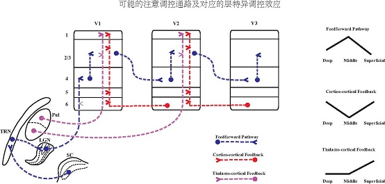
Fig 1. Possible neural pathways and its corresponding feedbacks of attention modulation
Researchers first collected high-resolution (0.75 mm isotropic) functional data with a T2* weighted 2D GE-EPI sequence, and T2* BOLD fMRI signals of V2 and V3. Using equal-volume method for depth calculation and spatial linear regression to mitigate volume effect, contract responses were obtained on the superficial, middle, and deep cortical depths. Results show additive effect from attention responses to modulation of T2* BOLD contrast response curves. Attention modulation was stronger in the superficial and deep cortical depths than in the middle depth of V1, consistent with the hypothesis that attention modulates early visual activity through direct cortico-cortical feedback connections. In V2 and V3, attention modulation was strongest in the superficial cortical depth, consistent with cortico-pulvino-cortical feedback to the superficial layers of extrastriate visual cortex. Experiments consistently showed that top-down spatial attention can modulate contrast responses in the superficial and deep layers of human early visual cortex, supporting the hypothesis that attention operates through feedback connections along descending visual pathway in a hierarchical fashion.
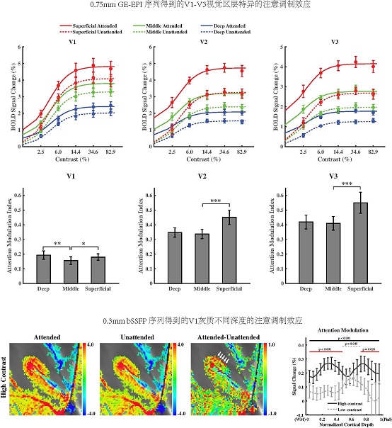
Fig 2. Attention modulation effect of V1, V2 and V3 regions of the visual cortex from the 0.75 mm GE-EPI sequence and attention modulation effect of different depths of V1 region from 0.3 mm bSSFP sequence
To further validate these findings, researchers used ultra-high resolution (0.3 mm in-plane) distortion free T2 weighted bSSFP fMRI method, and obtained T2 BOLD fMRI signals on varied depths of V1 gray matter. In view of high sensitivity to non-specific intravascular responses from large veins by T2* BOLD, T2 BOLD is more sensitive to microvasculature oxyhemoglobin concentration in the gray matter. Top-down spatial attention showed additive offset on T2* weighted GE BOLD but multiplicative scaling effect on T2 weighted bSSFP fMRI signals in the superficial and deep layers of human early visual cortex. Attention modulation of the low contrast response of bSSFP was also strongest in the middle cortical depths of V1. Researchers also used a BOLD biophysics model (Davis model) to verify that the additive shift of attention on T2* BOLD signal is due to the strong nonlinear relationship between BOLD signal of large blood vessels and underlying neural activity, while the responses from microvessels are relatively linear thus preserved the multiplicative scaling effect of neural modulations.
This research applied high-resolution fMRI at 7T to reveal the modulation mechanism of spatial attention on the contrast responses across different cortical depths in human early visual cortex, which has addressed a long-term discrepancy on attention modulating mechanism on bioelectric signals and T2* BOLD fMRI signals, from a perspective of MRI imaging principles. The role of high-resolution fMRI at ultra-high magnetic field in human brain and cognitive researches was highlighted. IBP postgraduate students - LIU Chengwen and GUO Fanhua - are co-first authors, and IBP researcher ZHANG Peng is correspondence author. IBP researcher HE Sheng contributed to design of cognitive experiment paradigm. Prof. Danny JJ Wang of Stevens Neuroimaging and Informatics Institute, University of Southern California provided instructions on the MRI physics. Dr. ZHANG Zihao operated the 7T MRI platform at IBP, and Dr. QIAN Chencan supported the MRI data processing. This work was supported by grant from Natural Science Foundation of China and Beijing Municipal Science & Technology Commission.
The web link for this research paper:: https://www.sciencedirect.com/science/article/pii/S0301008220301520
Contact: ZHANG Peng
Institute of Biophysics, Chinese Academy of Sciences
Beijing 100101, China
Email: zhangpeng@ibp.ac.cn
(Reported by Dr. ZHANG Peng's group)