
Scientists Develop Tetra-color superresolution microscopy based on excitation spectral demixing
The development of single-molecule localization microscopy (SMLM) provided a powerful tool for exploring nanostructures in cells that were previously concealed by diffraction limits. Besides the spatial resolution, multicolor imaging in SMLM is another important aspect, which is critical for investigating protein colocalizations and organelle interactions in biological research.
Recently, researchers from the Institute of Biophysics (IBP) of the Chinese Academy of Sciences (CAS) have developed a new multicolor SMLM method called excitation-resolved stochastic optical reconstruction microscopy (ExR-STORM). The work was published in Light Science & Applications online on Jan. 02, 2023.
This method utilized 3 excitation lasers (620 nm, 639 nm and 671 nm), which were synchronized with a resonant scanner. In this manner, the single molecule image under 3 excitation lasers could be obtained in one frame. "The resonant mirror is the key point in our method, since single molecules blink much faster than the exposure time of the camera, we cannot record the right photon number distribution without this fast-switching technique." As the researchers said.
To verify the capability of this method, microtubules, intermediate filaments, endoplasmic reticulum and the outer mitochondrial membrane were labeled with DyLight 633, Alexa Fluor 647, Dyomics 654 and CF660C, respectively. And tetra-color super-resolution images were reconstructed successfully, with negligible cross-talk. This method yields several advantages. First, dyes with similar emission spectra could be resolved, resulting in negligible chromatic aberrations. Second, it resolved 4 far-red dyes with only one objective, extending color channels with cross-talk of less than 3%. Third, the high scanning speed could eliminate the brightness fluctuation of single molecules, compatible with fast dynamics of STORM imaging.
In short, this research provides a powerful tool for high-throughput investigation of the 3D nanostructures in cells. This capability shows its great potential in the biological and medical research. The researchers believe that ExR-STORM will allow further investigation of organelle organization and interactions in cell stress responses, migration, division and differentiation.
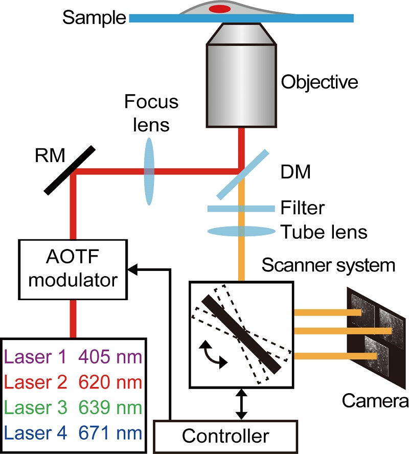
Figure 1 | Working principle of ExR-STORM.
Three excitation lasers (620 nm, 639 nm and 671 nm) and one activation laser (405 nm) were combined and then modulated by an AOTF prior to the illumination of the sample. The excitation laser switching was synchronized with the scanner, resulting in three subregions with different excitation wavelengths within one exposure time.
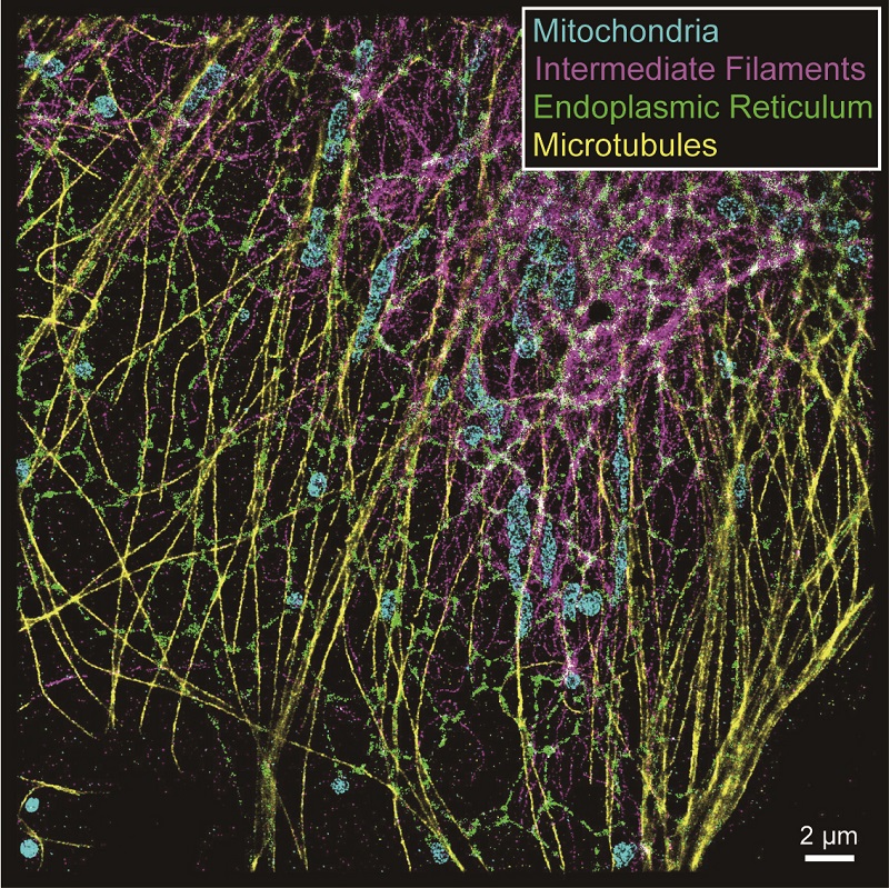
Figure 2 | Tetra-color imaging based on ExR-STORM.
CF660C, Alexa Fluor 647, Dyomics 654 and DyLight 633 were selected to label the outer mitochondrial membrane (cyan), intermediate filaments (magenta), endoplasmic reticulum (green), and microtubules (yellow), respectively, in a fixed COS-7 cell.
Article link: https://www.nature.com/articles/s41377-022-01054-6
Contact: JI Wei
Institute of Biophysics, Chinese Academy of Sciences
Beijing 100101, China
Email: jiwei@ibp.ac.cn
(Reported by Dr. JI Wei's group)