| Chinese Academy of Sciences Protein Science Core Facility Center |
|
Chinese Academy of
Sciences Protein
Science core facility
Center |
|
|
Titan Krios 300kV transmission electron microscope |
| Author: |
|
Update time: |
2014-11-26 |
|
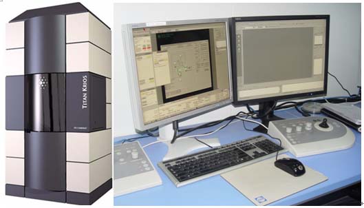
The Titan Krios™ transmission electron microscope (TEM) is tailored for use in protein and cellular imaging. Its revolutionary cryo-based technology and stability permit a full range of semi-automated applications, including 2D electron crystallography, single particle analysis, cryo electron microscopy, and dual-axis cellular tomography of frozen hydrated cell organelles and cells. The Titan Krios is the most powerful and flexible high resolution electron microscope for 3D characterization of biological samples, which has the point resolution of 0.27 nm and the limit of 0.14nm. The innovative enclosed platform combines excellent optical performance and thermal and mechanical stability with a high throughput sample loader. The Autoloader™ sample loader allows for the loading of up to 12 samples in a specially designed Autogrid™ ring. In addition, the specially designed holder allows the sample to rotate 90 degrees in plane while present in the column. This provides a reduction of the missing wedge information to a missing cone. The flexibility of operating the Titan Krios in the range of 80-300 kV allows for optimizing high tension to the requirements of the material under examination, ranging from vitrified suspensions to unstained cryo sections. The new digital user interface gives the freedom to operate the Titan Krios remotely in ambient, normal light conditions. The high-speed digital camera (Smart cam), which has taken over the role of the fluorescent screen, and the innovative user interface improve the microscope’s ease of use. It masters the complete dynamic range from live observation of focused high intensity beams to low dose applications and diffraction. The instrument is used for high resolution 3-D structure study of biomacromolecule complex, and high resolution 3-D ultrastructure imaging for cell (organells). Technical Support: Gang Ji (010-64888419, jigang@moon.ibp.ac.cn) Xiaojun Huang (010-64888419, xiaojunhuang@moon.ibp.ac.cn) |
|
|
|